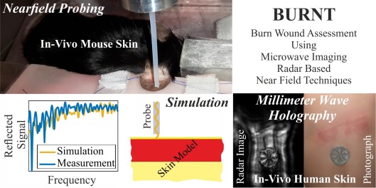
Burn Wound Assessment Using Microwave Imaging Radar Based Near Field Techniques
Phase 1 and 2
In modern burn surgery the fast and precise assessment of the burn depth is challenging but also of high importance to adapt the correct wound care. For this purpose this research project is to systematically investigate and develop methods of contactless millimeter wave and terahertz imaging techniques for diagnosing burns.
Participating Institutes and Institutions:
Abteilung für Plastische Chirurgie und Schwerbrandverletzte, Handchirurgiezentrum, Berufsgenossenschaftliches Universitätsklinikum Bergmannsheil, Ruhr-Universität Bochum
Lehrstuhl für Hochfrequenztechnik, Friedrich-Alexander-Universität Erlangen-Nürnberg
Research Team:
Prof. Dr.-Ing. Martin Vossiek
PD Dr. med. Ole Goertz
Prof. Dr.-Ing. Dr.-Ing. habil. Helmut Ermert
Dipl.-Ing Julian Adametz
M.Sc. Daniel Oppelt
Kristina Zhuravleva
Objectives
The purpose of this research project is to systematically investigate and develop, for the first time, the full potential of terahertz (THz) imaging techniques for diagnosing burns with non-contact, fully coherent, reconstructive THz imaging techniques for medical diagnosis. In the course of the project two alternative approaches will be investigated and compared: (1) tomographic radar imaging with a multiple-input-multiple-output (MIMO) radar concept, and (2) a near field scanner with non-contact probes for assessing the evanescent near field. Both approaches will be experimentally tested on a common test platform.
The tomographic radar imaging approach will focus on a fully coherent, holographic technique. A multistatic MIMO imaging array configuration for the application of THz waves up to 500 GHz based on a sparse periodic array setup will be developed. A coherent THz measurement system, employing two antenna elements, two independent positioning units for the Rx and Tx antenna, and a control computer will be developed and manufactured. This setup will allow testing arbitrary Rx/Tx MIMO arrangements by sequential measurements. This is the most cost efficient and the most flexible way for experiments. As measuring speed is not an issue in a fundamental research state, the long duration of the sequential measurement procedure is of minor importance. Along with smart THz sensor system concepts, sophisticated reconstruction methods will be investigated, which deliver a high-resolution 3D image of burned skin regions and a highly improved diagnostic reports on burn depth and blood perfusion compared to the state of the art. The whole radar system will be designed in a polarization sensitive manner. This means, that the system will be able to measure the entire scattering behavior of samples in terms of the vertical-horizontal, horizontal-vertical, vertical-vertical and horizontal-horizontal components. Fundamental research will be carried out towards novel approaches for tissue classification by means of a fully polarimetric analysis.
Furthermore, a THz near field microscopy imaging system will be designed, implemented and examined for its capability in burn wound assessment. For this purpose, a dielectric near field probe to access and measure the evanescent near field scattered by burned skin areas will be constructed first. The combination of several probes to form an array structure will be examined subsequently. A highly precise distance control system to ensure the detection of the near field and prohibiting unpleasant contact between the probing elements and the skin will be developed. A novel image reconstruction algorithm will be implemented to account for both near field propagation effects and biological tissue scattering phenomena. For the generated images of the skin, an outstandingly high resolution capability is expected, allowing for a vastly enhanced diagnosis of the severity of burn wounds. The potential of the combination of results of both near field microscopy and radar tomography will be investigated.
The suitability of the two proposed concepts for clinical diagnostics will be verified by experimental tests on phantom materials as well as in-vivo examinations of burns.
Abstract
Large burns are among the worst injuries suffered by people – they often have life-threatening effects and serious negative impacts on the quality of life. Diagnosing and treating burns is a very complex procedure. The key to successful wound treatment is to quickly estimate the depth of the burn. This is challenging and requires a lot of experience on the part of the medical practitioner. One of the main reasons why current diagnostic procedures are so complex is that no machine systems for imaging burns for clinical assessment are available that have proven successful in clinical pathology.
Among the approaches available to date for burn diagnosis are laser Doppler imaging (LDI) and optical coherence tomography (OCT). THz imaging techniques have been introduced which bring the additional benefit of passing through dressings which is not possible with OCT and LDI. All THz systems available to date are fairly basic incoherent scanners with strongly-focused antennas or tactile probes. As with LDI and OCT the skin is slowly scanned one position at a time.
The purpose of this research project is to systematically investigate and develop, for the first time, the full potential of terahertz (THz) imaging techniques for diagnosing burns with non-contact, fully coherent, reconstructive THz imaging techniques for burn diagnosis. In the course of the project two alternative approaches will be investigated and compared: (1) tomographic radar imaging with a multiple-input-multiple-output (MIMO)-based data logging system, and (2) an array of non-contact probes for assessing the evanescent near field. Along with smart THz sensor system concepts, reconstruction methods will be investigated, which deliver a high-resolution 3D image of the burn and much improved diagnostic reports on burn depth and blood perfusion. Fundamental research will be carried out into novel approaches to tissue classification by means of polarization sensitive analysis. The suitability of these concepts for clinical diagnostics will be verified by experimental tests on phantom materials as well as in-vivo examinations of burns.
This joint interdisciplinary project combines the expertise of the Institute of Microwaves and Photonics (LHFT) at Erlangen University in Germany – one of the leading research centers worldwide for coherent reconstructive wireless imaging systems, and the Clinic for Plastic Surgery and Severe Burn Injuries at the Ruhr University Bochum, Germany – a nationally and internationally renowned clinic in this field. The goal of the joint endeavor is to investigate next-generation approaches for burn diagnostics, with the hope of making the long-awaited breakthrough in the long-term.
Publications
D. Oppelt, T. Pfahler, F. Distler, J. Ringel, O. Goertz and M. Vossiek, “Nearfield Imaging Probe for Contactless Assessment of Burned Skin – to be published,” in Proceedings of the 47th European Microwave Conference (EuMC), Nuremberg, Germany, Oct. 2017.
D. Oppelt, J. Adametz, J. Groh, O. Goertz and M. Vossiek, “MIMO-SAR Based Millimeter-Wave Imaging for Contactless Assessment of Burned Skin,” in Proceedings of the International Microwave Symposium (IMS2017), Honolulu, Hawaii, USA, June 2017.


